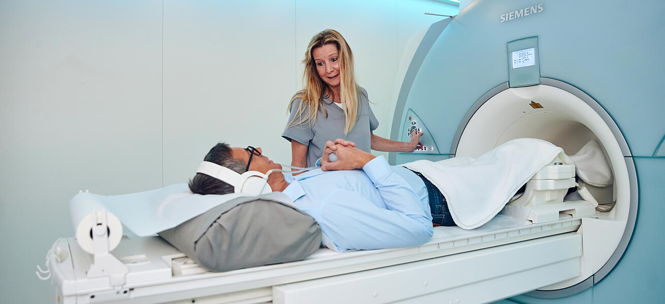
MRI (Magnetic Resonance Imaging)
MRI Munich – a brief introduction to how magnetic resonance imaging works
Magnetic resonance imaging, or MRI, is an important diagnostic procedure used to visualize the inside of our bodies. It is an imaging technique that can be used to visualize the structures and functions of tissues and organs.
Magnetic resonance imaging was discovered as early as the 1950s. However, it was first used mainly in the fields of chemistry and physics. It was not until the mid-1990s that it was also used in medicine.
Magnetic resonance imaging – a brief introduction to how it works
Magnetic resonance tomographs do not work with X-rays, but use special magnetic fields. In our body there are atomic nuclei, which usually rotate around themselves. The name nuclear spin is also derived from this phenomenon. Magnetic resonance imaging now generates a magnetic field that lies outside the body. Thus, the atomic bodies are forced to arrange themselves in a certain direction.
But not only is a magnetic field generated in an MRI scanner, but radio waves are also directed at the body. The radio waves are delivered to the body at specific intervals. During the pauses, signals are emitted by the atomic nuclei. This allows an accurate picture of your interior to be mapped. For certain examination procedures, it may be necessary to inject contrast medium intravenously during the examination. This contrast agent is distributed throughout the body by intravenous administration. This makes it easier to distinguish between different tissues and enables meaningful diagnostics.
With an MRI, the entire body can be scanned, so to speak, or only certain areas can be scanned.


