The diagnosis calcific shoulder initially worries many patients about surgery. But we can reassure you: modern orthopaedics now offers a wide range of highly effective, gentle treatments that make surgery unnecessary in most cases. As an experienced orthopaedic center in Munich, we support you on your way back to a pain-free, mobile everyday life – without the burden of a surgical procedure.
Although the pain associated with a calcific shoulder can be extremely severe and make everyday life a struggle, it is fundamentally a condition with a strong tendency to heal on its own. The key to success is to specifically support the body in its natural breakdown processes and to significantly shorten the painful phases. Modern treatments such as shock wave therapy have proven themselves here and can support healing without surgery. In this comprehensive guide you will learn everything about the leading conservative treatment approaches, how they work and how calcific shoulder treatment without surgery can help you become pain-free again.
At a glance
The key points in brief
- Calcific shoulder is a common condition of the rotator cuff and can be treated without surgery in most cases.
- Typical are sudden, severe pain, especially at night and during overhead movements.
- Ultrasound and X-ray show the location, size and consistency of the calcium deposit.
- Shock wave therapy is a central component of conservative treatment.
- Surgery only becomes an option if conservative treatment has not helped sufficiently over several months.
Table of contents:
- What is a calcific shoulder and how does it develop?
- Anatomy of the rotator cuff
- Symptoms and diagnosis – modern imaging
- Shock wave therapy – gentle and without surgery
- Other conservative treatments
- Physiotherapy and home exercises
- When is surgery really necessary?
- Prognosis and course of healing
- Frequently asked questions (FAQ)
- Conclusion – modern orthopaedics relies on gentle methods
What is a calcific shoulder and how does it develop?
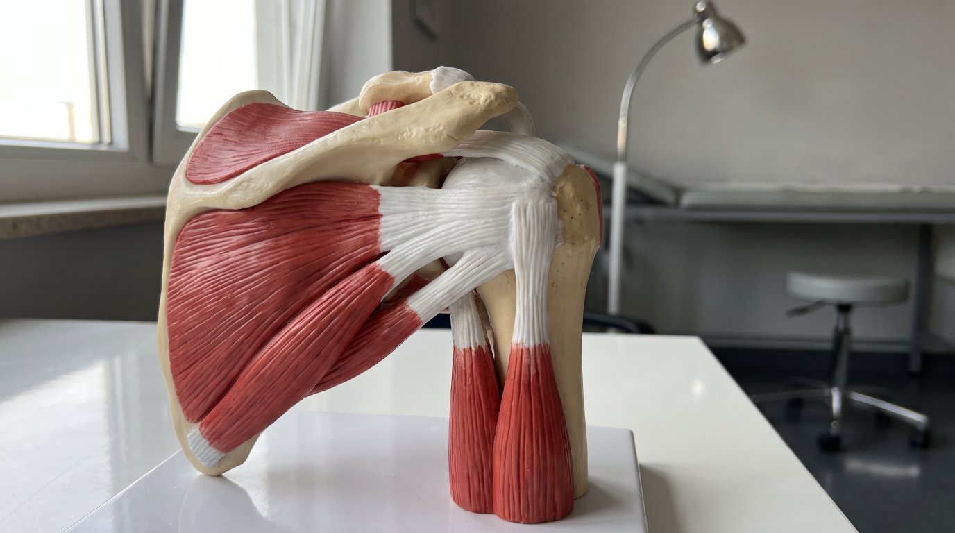
The tendinosis calcarea, commonly known as calcific shoulder, is a condition of the rotator cuff in which calcium deposits form in the shoulder tendons. These deposits are not caused by mechanical wear or overuse, but follow a characteristic biological process that unfolds in three clearly defined phases and can extend over months to years.
The three-phase model of calcific shoulder is central to understanding the condition: in the first phase, precalcification, normal tendon cells transform into cartilage cells through mechanisms that are not yet fully understood. These cellular changes create the conditions for the calcium deposits that follow. During the second phase, calcification, chalky, firm calcium deposits form first and may later take on a softer, toothpaste-like consistency. The third and often most painful phase, postcalcification or resorption phase, marks the body’s own breakdown process. Here the immune system becomes active, scavenger cells migrate in and begin to dissolve and carry away the calcium deposits. This natural cycle underlines the body’s fundamental ability to heal itself, even though without treatment this process can be very lengthy and painful.
Who is affected? Calcific shoulder is a common shoulder condition. Women are affected more often than men, and incidence peaks between the ages of 40 and 60, a stage of life in which work and family demands are often particularly high. The calcium deposits most commonly form in the supraspinatus tendon, less often in the infraspinatus or subscapularis tendon. This distribution is related to the particular anatomy of the different tendon sections.
The causes of calcific shoulder have not yet been fully clarified. Contrary to what is often assumed, it does not result from overuse or degenerative wear. Instead, local circulatory disturbances, genetic factors and disorders of calcium metabolism appear to play a role. The fact that women are affected more often also suggests hormonal influences. It is important to know that calcific shoulder is not caused by a poor diet or too much calcium – these widespread assumptions are not medically tenable.
1PrecalcificationPhase 1
Tendon cells change – usually without symptoms at this stage.
2CalcificationPhase 2
Calcium deposits form, firm at first and softer later.
3ResorptionPhase 3
The body breaks down the calcium – often the most painful phase.
Anatomy of the rotator cuff
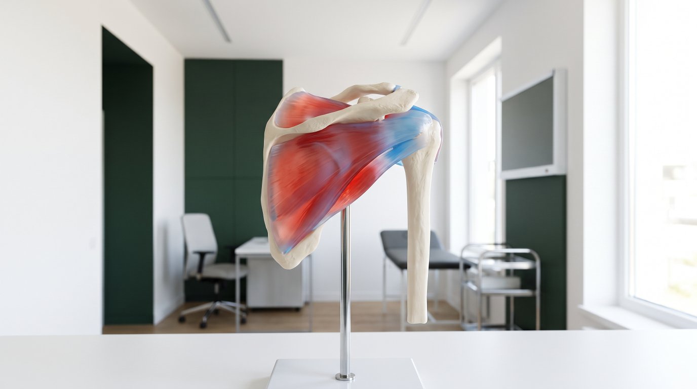
The shoulder is the most mobile ball-and-socket joint in the human body and allows movement in all directions – a property that makes it unique. This exceptional mobility is ensured by a complex interplay of bones, ligaments, muscles and tendons. A central role is played by the rotator cuff, a group of four muscles that run from the shoulder blade to the head of the upper arm and enclose it like a protective cap.
The four muscles of the rotator cuff each have specific functions and work together harmoniously: the supraspinatus muscle is mainly responsible for lifting the arm out to the side and for centering the humeral head in the socket. The infraspinatus muscle and the teres minor muscle are responsible for external rotation of the arm, while the subscapularis muscle enables internal rotation. Their tendons fuse into a common tendon plate that envelops the humeral head and provides both stability and the complex movement patterns of the shoulder.
The supraspinatus muscle holds a special position, because its tendon runs through the anatomically narrow subacromial space between the humeral head and the shoulder roof (acromion). This area is also known as the “critical zone”, because blood supply here is naturally reduced. The reduced oxygen supply makes this region particularly susceptible to degenerative changes and the formation of calcium deposits. In addition, movements can cause mechanical narrowing that aggravates the problem. These anatomical features explain why the supraspinatus tendon in particular is so often affected by calcium deposits and why targeted treatment is so important to avoid complications such as a stiff shoulder.
Symptoms and diagnosis – modern imaging
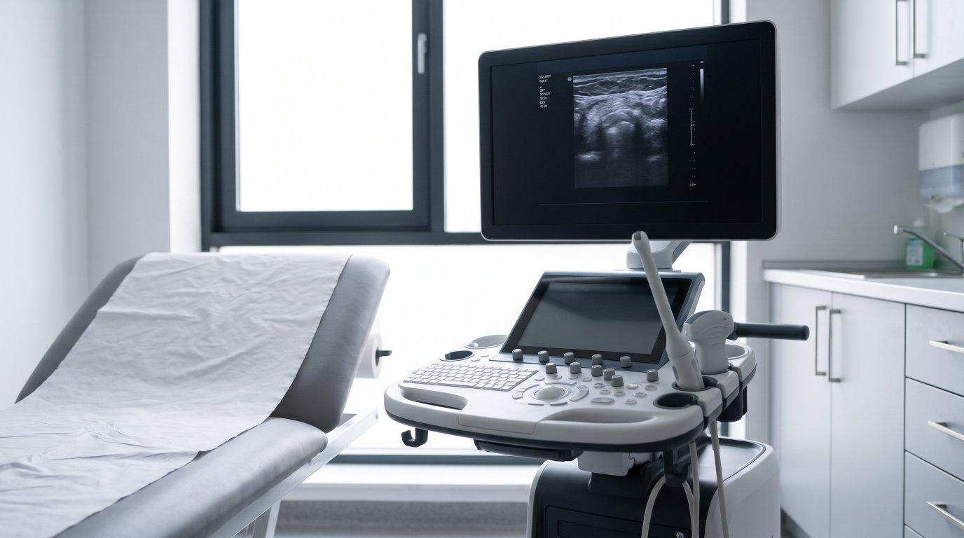
The symptoms of a calcific shoulder are often described among specialists as the “chameleon of shoulder pathologies”, because the range of symptoms can be extraordinarily varied. This variability makes the condition a diagnostic challenge for patients and doctors alike. While some people feel no symptoms at all and the calcium deposits are only discovered by chance during other examinations, others suffer unbearable pain that can make life a torment.
Typical symptoms include suddenly occurring, severe pain that often appears without an identifiable trigger and becomes more intense at night in particular. This pain at rest at night makes it impossible to lie on the affected side and leads to considerable sleep disturbances. During the day, load-dependent pain occurs mainly with overhead movements – putting on a sweater, combing your hair or reaching for objects on shelves becomes a painful ordeal. The symptoms often radiate from the shoulder into the upper arm and can be accompanied by marked restrictions of movement. The pain is particularly intense during the resorption phase, when the immune system begins to actively break down the calcium deposits and triggers a massive inflammatory reaction in the surrounding tissue.
The modern diagnosis of a calcific shoulder relies on a combination of clinical examination and imaging. As experienced orthopaedists, we can already gain important clues for the diagnosis through targeted function tests and range-of-motion assessments. For definitive confirmation and precise characterization of the calcium deposits, however, imaging is indispensable:
- Ultrasound (sonography): Today regarded as the gold standard in calcific shoulder diagnostics. Modern high-resolution ultrasound devices allow a dynamic examination and show the exact location, size and consistency of the calcium deposit as well as any accompanying bursitis with precision.
- X-ray: Makes the calcium deposit visible as a characteristic dense shadow and allows its size and shape to be assessed. Two views (anterior-posterior and lateral) are required for a complete picture.
- Magnetic resonance imaging (MRI): Needed less often, but can provide valuable additional information in unclear findings or to distinguish calcific shoulder from tendon tears and other complex shoulder pathologies.
However, the clinical assessment always takes precedence over imaging alone. What matters for our treatment planning is not just the size of the calcium deposits or how they look on the X-ray, but above all the symptoms and the individual level of suffering of the patient. A precise distinction from other shoulder conditions such as impingement syndrome, frozen shoulder or tendon tears is very important, as these require different treatment approaches.
Shock wave therapy – gentle and without surgery
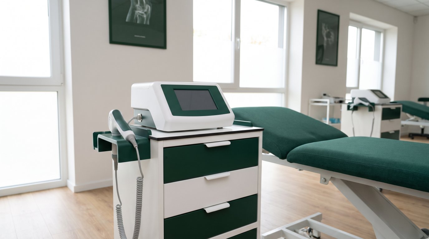
Among the conservative treatment methods, extracorporeal shock wave therapy (ESWT) has become established as an important non-surgical option. This non-invasive form of treatment has been used in orthopaedics since the end of the 1980s and can be an alternative to surgery for many patients. At the Orthopaedic Center Munich East, shock wave therapy is an integral part of our range of treatments.
The mechanism of action of shock wave therapy is multifaceted: the high-energy sound waves are directed from outside precisely at the calcium deposit in the shoulder tendon, where they take effect. Mechanically, the shock waves fragment the calcium crystals and make them accessible to the body’s own breakdown processes. At the same time, blood flow in the treated tissue is significantly increased and tendon metabolism is activated. This speeds up both the removal of the calcium fragments via the lymphatic system and the regeneration of the damaged tendon tissue. In addition, the body’s own growth factors are released, which further promote the healing process and stimulate the formation of healthy replacement tissue.
Studies show that shock wave therapy can relieve pain and improve shoulder function in calcific shoulder. How well it works in an individual case depends, among other things, on the size, location and consistency of the calcium deposit. Starting treatment early generally has a favorable effect.
The course of treatment usually comprises several sessions about a week apart; we determine the number individually. Treatment usually takes only a few minutes and is carried out on an outpatient basis. Beforehand, the calcium deposit is located by imaging so that the shock waves reach exactly the right spot. Many patients notice an improvement during the series, but the full effect often only shows a few weeks after treatment has finished.
Focused vs. radial shock wave therapy
Modern shock wave therapy distinguishes between two different forms of application that differ in their physical mode of action and penetration depth: the focused and the radial shock wave. Both methods have their specific strengths and can be used individually or in combination depending on the findings.
Focused shock waves concentrate their energy precisely deep in the tissue and can be aimed at the calcium deposit with millimeter accuracy. They act with high energy and in a targeted way, which makes them particularly effective for directly fragmenting the calcium deposits. The radial shock wave, by contrast, releases its energy more superficially and spreads out in a fan shape through the tissue. It is excellent for treating muscular tension and for loosening the surrounding tense tendon and fascial tissue, which is often badly affected by inflammation and pain-related protective posture.
A combination of both methods can make sense. It allows us to address not only the calcium deposit itself but also to treat the entire functional environment of the shoulder. While the focused shock wave directly fragments the calcium deposit, the radial shock wave treats the surrounding muscle and tendon structures. Whether this combination is right for you, we decide after the examination.
Orthopaedic Center Munich East
Have your shoulder pain assessed by a specialist
We examine your shoulder thoroughly, including by ultrasound, and discuss the right treatment with you.
Other conservative treatments
In addition to shock wave therapy, other proven conservative treatment options are available, which we use or combine in a targeted way depending on the phase of the disease and the individual situation. Phase-appropriate treatment takes into account the current stage of the condition and the patient’s present symptoms.
Drug-based pain therapy with non-steroidal anti-inflammatory drugs (NSAIDs) is often the first line of treatment, especially in acute pain phases. These medications, such as ibuprofen, diclofenac or etoricoxib, relieve pain and reduce inflammation and can significantly ease the often unbearable pain of the resorption phase. In cases of very severe pain, stronger analgesics may be needed for a short time, and we always aim for balanced pain therapy with as few side effects as possible.
Injection treatments are another important therapy option that may be considered depending on the symptoms. Cortisone injections under the shoulder roof (subacromial infiltrations) can bring rapid pain relief during acute inflammatory phases. The injection is placed precisely in the inflamed bursa and acts directly at the site of inflammation. We use cortisone sparingly and only in limited numbers.
Physical therapy measures can usefully complement treatment
- Cold therapy: In acute inflammatory phases for pain relief and to reduce inflammation
- Heat therapy: In chronic cases to promote blood flow and muscle relaxation
- Electrotherapy and ultrasound therapy: For pain modulation and tissue activation
- Electromagnetic transduction therapy (EMTT): As a supportive measure to promote regeneration
Patience and consistency are of central importance in conservative treatment. We always tell our patients honestly that courses of treatment lasting several months are completely normal and should in no way be interpreted as treatment failure. Combining different approaches – in particular shock wave therapy with physiotherapy and, where needed, medication – optimizes the chances of success and in most cases leads to a significant improvement without surgery.
Good to know: A calcific shoulder is not caused by too much calcium in the diet. A low-calcium diet therefore does not help and is even unfavorable for your bones.
Physiotherapy and home exercises

Physiotherapy plays a central and indispensable role in the conservative treatment of calcific shoulder. We prescribe the appropriate physiotherapy and tailor its focus to your findings. The main goal of physiotherapeutic treatment is to maintain and improve the mobility of the shoulder joint while preventing pain-related stiffening (frozen shoulder) – a feared complication that can considerably delay healing.
Special capsule stretching exercises are at the heart of treatment. In painful conditions the joint capsule of the shoulder tends to shorten and thicken, which leads to considerable restrictions of movement. Targeted stretching exercises in different directions – especially external rotation, internal rotation and elevation – can prevent or reverse this shortening. These exercises are first performed passively by the therapist, later actively assisted and finally actively by the patient.
Strengthening the rotator cuff muscles is another important building block of therapy. Pain-related protective posture weakens the stabilizing muscles of the shoulder, which can lead to imbalances and further problems. A systematic strengthening program that starts with light resistance and is increased step by step helps restore normal shoulder function and prevent further overload. It is important that the exercises are adapted to pain – they should challenge but not cause additional damage.
Posture training and ergonomic advice often address existing incorrect loading that may have contributed to the development of the calcific shoulder. Many of our patients suffer from posture-related problems caused by screen work, one-sided strain or unfavorable sleeping positions. Targeted education and practical exercises can correct these problems.
The patient’s own responsibility is of decisive importance for the success of treatment. Home exercises performed regularly and correctly can speed up healing considerably and effectively prevent relapses. A home exercise program that you learn in physiotherapy helps you actively work on your recovery between therapy appointments as well.
When is surgery really necessary?
Surgery for calcific shoulder should always be the exception and by no means the rule. As a modern orthopaedic center, we consistently follow the principle: “As conservative as possible, as surgical as necessary.” This philosophy has proven its worth not only from a medical point of view, but also meets our patients’ natural wish for gentle, low-risk treatment.
The clear indication for a surgical procedure requires several criteria to be met at the same time: conservative treatment should generally have been carried out consistently over several months (often cited is about six months) without achieving sufficient pain relief or functional improvement. All available conservative options – in particular shock wave therapy, physiotherapy and injection treatments – must have been applied properly. In addition, there must be treatment-resistant pain in an otherwise healthy patient that significantly impairs quality of life and causes marked functional limitations in everyday life.
Prerequisites for a surgical intervention also include an appropriate balance between surgical risk and expected benefit. Here we take into account the patient’s age, existing conditions, occupational demands and personal expectations. Realistic information about the surgical risks, the expected course of healing and the necessary follow-up care is essential. Patients must understand that even after surgery several months of rehabilitation are required and that complete freedom from symptoms cannot be guaranteed in every case.
Minimally invasive arthroscopic procedures are now regarded as the gold standard of surgical calcific shoulder treatment when an operation is truly unavoidable. This modern keyhole technique allows precise removal of the calcium with minimal trauma to the surrounding tissue. Through a few small skin incisions, a mini camera and fine instruments are introduced into the shoulder joint to locate and remove the calcium deposit under direct vision. At the same time, any accompanying pathologies such as bursitis or small tendon tears can be treated as well.
Second opinions before surgical procedures are generally recommended and are expressly welcomed by us as reputable practitioners. An independent specialist assessment ensures that all conservative options have really been exhausted and that the operation is truly medically indicated. We therefore actively encourage our patients to seek another orthopaedic opinion if they are unsure – because only a well-informed patient can make a sound decision.
Prognosis and course of healing
Calcific shoulder is fundamentally a self-limiting condition with a very good long-term prognosis. This means the human body has the natural ability to break down and reabsorb the calcium deposits on its own. This biological fact gives us the certainty that healing is in principle always possible – even though without targeted treatment this self-healing process can take months to years and is associated with considerable pain.
Factors for a favorable prognosis are manifold and in part within the patient’s control: starting treatment early, before chronic pain cycles have become established, clearly improves the chances of success. Consistently carrying out the prescribed treatments – especially shock wave therapy and physiotherapy – is decisive for success. The patient’s willingness to take an active part, including regularly doing home exercises and observing ergonomic principles in everyday life, can speed up healing considerably. Age also plays a role – younger patients often heal faster because their tissues regenerate better.
The individual course, however, varies greatly and depends on several factors: the size and consistency of the calcium deposit influence both the intensity of the symptoms and the time until it dissolves completely. The stage of the condition at the start of treatment also matters – patients in the early calcification phase often respond faster to treatment than those in the late resorption phase. Physical constitution, blood supply and immune system activity vary individually and have a decisive influence on healing.
Realistic expectations of timing help our patients go through treatment calmly. First improvements may be noticeable after just a few weeks, but complete freedom from pain usually only occurs after three to six months. The radiologically detectable dissolution of the calcium deposits can take even longer and often only follows the clinical improvement. It is important to understand that fluctuations during healing are normal and should not be seen as a setback.
Follow-up care includes regular check-ups at our center to monitor healing progress and adjust the treatment strategy if needed. Continuing movement-preserving measures and preventive exercises even after the acute symptoms have subsided is important to avoid relapses. The risk of recurrence is low with proper treatment and can be reduced further through preventive measures such as regular shoulder exercises, ergonomic workplace design and avoiding one-sided strain.
Important: Courses lasting several months are normal in calcific shoulder. Fluctuations during healing are not a sign that the treatment is not working.
Orthopaedic Center Munich East
Your appointment at OZMO
Do you have shoulder pain, or have you already been advised to have surgery? Make an appointment – a second opinion is also possible on request.

Frequently asked questions (FAQ)
How long does it take for calcium in the shoulder to dissolve?
The time it takes for calcium deposits to dissolve is highly individual and can range from a few months to several years. Without treatment, this natural process can take two to four years. The duration depends on several factors: the size and consistency of the calcium deposit, the stage of the condition, the activity of the body’s own immune system and the chosen treatment. Targeted calcific shoulder treatment without surgery, in particular with shock wave therapy, can speed up this process considerably. Many patients experience significant pain relief after just a few weeks, although complete dissolution of the calcium deposits can take several months.
How many sessions of shock wave therapy are needed for calcific shoulder?
Extracorporeal shock wave therapy usually involves several sessions, often three to five, about a week apart. After the first sessions we check together how the treatment is responding. The exact number of treatments needed depends on your individual response to therapy, the size and consistency of the calcium deposit and how the pain develops. Many patients feel a clear improvement after the first or second treatment, but the full treatment success with dissolution of the calcium deposit usually only appears after the entire treatment series is complete. For particularly large or stubborn calcium deposits, additional sessions can make sense.
Can a calcific shoulder dissolve on its own?
Yes, calcific shoulder is fundamentally a self-limiting condition with a high tendency to heal spontaneously. The human body has its own immunological mechanisms that are able to completely break down and reabsorb the calcium deposits. However, this natural course can be very lengthy and drag on for several years. During this time, patients are often exposed to recurring phases of severe pain that can considerably impair quality of life. The goal of conservative treatment is therefore not to force a process that would not take place anyway, but to specifically accelerate the body’s own healing, significantly shorten the painful phases and prevent complications such as a stiff shoulder.
Is a calcific shoulder caused by too much calcium in the diet?
No, this widespread assumption is not medically tenable. The development of a calcific shoulder has nothing to do with calcium intake from food. The calcium deposits arise from local cell changes in the tendon itself, which cause tendon cells to transform into calcium-producing cells. A low-calcium diet therefore does not help in treatment and can even be harmful, as calcium is essential for bone health. Instead, it is important to concentrate on proven treatment methods.
Is sport possible with a calcific shoulder?
Sport is generally possible and often even beneficial for healing, but the activity should be adapted to the current pain phase. During acute pain phases, overhead movements and strenuous shoulder loading should be avoided. Cycling, walking or jogging are usually well suited. Swimming is possible as long as the arm movements are pain-free. We will discuss with you individually which load makes sense at the moment. It is important that sport is never done through severe pain, as this can aggravate the inflammation.
How does a calcific shoulder differ from other shoulder conditions?
Calcific shoulder differs from other shoulder conditions in several characteristic features: the pain often appears suddenly and without an identifiable trigger, is typically more intense at night and radiates into the upper arm. Unlike in impingement syndrome, the symptoms are very severe in phases but can then disappear completely. The definitive distinction is made by imaging, in particular ultrasound and X-ray, which make the characteristic calcium deposits visible. A specialist examination is essential for a reliable differential diagnosis.
Conclusion – modern orthopaedics relies on gentle methods
Calcific shoulder treatment without surgery is a proven therapeutic approach: in most cases a calcific shoulder can be treated in a targeted way and without surgery. A precise diagnosis, phase-appropriate treatment and a little patience are what matter.
Shock wave therapy is a central component. It can relieve pain, improve function and be an alternative to surgery for many patients. Complemented by individually tailored physiotherapy and supportive measures as needed, the result is a treatment plan that fits your situation.
Modern orthopaedics respects the body’s natural healing processes and supports them optimally rather than disturbing them with invasive procedures. Patience and trust in the body’s self-healing powers, combined with the possibilities of modern medicine, lead to the best result for our patients. We take the time for thorough information and advice, because only a well-informed patient can actively contribute to their recovery.
Our conclusion: Early orthopaedic advice and consistent conservative treatment are the keys to successful therapy. Do not hesitate to see a specialist if you have shoulder pain – the sooner treatment begins, the sooner you can return to a pain-free, mobile everyday life. At the Orthopaedic Center Munich East we are at your side with modern diagnostics and gentle treatment methods.


Tailored diagnostics and treatment – at three locations in the east of Munich. For all insured and private patients.
Memberships & partners



Part of the Ortivity Network · Association doctors of the German Ice Hockey Federation
Clinic Munich
Haar
Münchener Straße 14
85540 Haar
+49 (0) 89 4141 434 – 64
Fax +49 (0) 89 45 45 49 – 39
Opening hours
| Morning | Afternoon | |
|---|---|---|
| Mon | 08:30 – 12:00 | 14:00 – 18:00 |
| Tue | 08:30 – 12:00 | – |
| Wed | 08:30 – 13:00 | – |
| Thu | 08:30 – 12:00 | 14:00 – 18:00 |
| Fri | 08:30 – 12:00 | – |
Appointments by arrangement only
Clinic Munich
Bogenhausen
Denninger Straße 15
81679 Munich
+49 (0) 89 4141 434 – 64
Fax +49 (0) 89 45 45 49 – 39
Opening hours
| Morning | Afternoon | |
|---|---|---|
| Mon | 08:30 – 12:00 | 14:00 – 18:00 |
| Tue | 08:30 – 12:00 | – |
| Wed | – | – |
| Thu | 08:30 – 12:00 | 14:00 – 18:00 |
| Fri | 08:00 – 12:00 | – |
Appointments by arrangement only
Clinic Munich
Feldkirchen
Sonnenstraße 26b
85622 Feldkirchen
+49 (0) 89 90 31 21 4
Fax +49 (0) 89 90 31 52 4
Opening hours
| Morning | Afternoon | |
|---|---|---|
| Mon | 08:00 – 12:00 | 14:00 – 17:00 |
| Tue | 08:00 – 12:00 | – |
| Wed | 08:00 – 12:00 | – |
| Thu | 08:00 – 12:00 | 14:00 – 17:00 |
| Fri | 08:00 – 12:00 | – |
Appointments by arrangement only