Shoulder pain is one of the most common orthopedic complaints and severely limits daily life, work, and athletic activities. In this stressful situation, many people ask themselves anxious questions such as, “Why does my shoulder hurt when I lift my arm?” or “Could a torn tendon be causing my symptoms?” Anyone suffering from persistent shoulder pain wants, above all, quick clarity about the exact cause of their symptoms and a realistic assessment of possible treatment options before deciding on a specific course of therapy.
This article explains in clear and detailed terms the various causes of shoulder pain, how the precise diagnostic process works at the Orthopedic Center Munich East, and which conservative and surgical treatment options may be appropriate based on your individual findings. The goal is to provide you with a well-founded overview so that you can take the path toward a pain-free shoulder in an informed and self-directed manner. Both the expertise of Prof. Dr. Peter Diehl and the latest medical findings are incorporated into this guide to offer you a holistic perspective on the topic. This will equip you with a solid foundation of knowledge before you, if necessary, schedule an appointment with a specialist and begin the process of individually assessing your symptoms.

Prof. Dr. med. habil. Peter Diehl ist Facharzt für Orthopädie und Unfallchirurgie am OZMO – Orthopädie Zentrum München Ost. Als Spezialist für Schulter- und Knieerkrankungen verfügt er über langjährige operative Erfahrung. Zu seinen Behandlungsschwerpunkten zählen Sportverletzungen, Knorpelschäden, regenerative Knorpeltherapien und minimalinvasive Gelenkeingriffe.
Table of contents:
- Why Does My Shoulder Hurt? An Overview of Possible Causes
- Shoulder Impingement Syndrome
- Calcific tendinitis and calcium deposits
- Torn Tendon in the Shoulder and Rotator Cuff
- Diagnostics: How the Cause Is Identified
- Conservative Treatment Options
- When Arthroscopy Is Recommended
- Recovery After Tendon Surgery
- The Role of Physical Therapy
- What You Can Do for Your Shoulder
- When You Should See a Shoulder Specialist
- Frequently asked questions
- Conclusion


Why Does My Shoulder Hurt? An Overview of Possible Causes
Shoulder pain does not automatically have a clear, easily identifiable cause. Rather, for affected patients, there are always various—and sometimes overlapping—triggers to consider, which may be similar or significantly different in terms of symptoms and progression. Prof. Dr. Peter Diehl explains that in cases of sudden-onset shoulder pain, the bursa often becomes inflamed first—simply put, this is a kind of gel-filled cushion between the acromion and the tendons that normally ensures smooth movement.
It is important to clarify a widespread misconception: Bursitis is not necessarily the actual cause of the symptoms, but may rather be a noticeable symptom of an underlying problem. Only a thorough examination by an experienced specialist can determine which factor is actually responsible.
Possible causes of shoulder pain:
- Shoulder impingement or impingement syndrome
- Calcium deposits or calcifications in tissue
- Signs of wear and tear in the joint structures
- Shortening of surrounding muscles and tendons
- Tendon tears in the area of the joint capsule
Distinguishing Between Symptoms and Causes
To properly understand the complex causes of shoulder pain, one must take a nuanced look at the exact relationships involved. Instead of the blanket statement “Bursitis is the cause of shoulder pain,” it is more accurate from a medical standpoint to say: An inflamed bursa can directly cause pain. However, the key to long-term treatment success always lies in determining why the irritation or inflammation developed in the first place. This distinction between symptom and underlying cause characterizes the typical approach at the Orthopedic Center Munich East and forms the basis for every well-founded diagnosis.
A Structured Approach to Identifying the Cause:
- Recognize a symptom
- Determine the possible cause
- Examine Shoulder Function
- Assessing Structural Changes
- Develop a personalized treatment plan
Shoulder Impingement Syndrome: When the space under the acromion becomes narrow
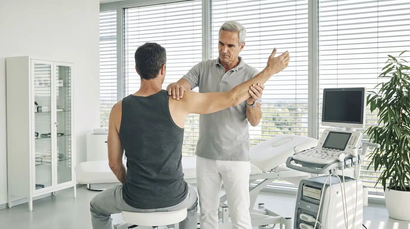
When raising the arm to the side, many patients may suddenly experience unpleasant, stabbing pain, which typically occurs when structures beneath the acromion are mechanically constricted. Tendons and a bursa run through this confined space—if this area becomes too narrow, the bursa can become inflamed and the tendon can be irritated by constant friction. Those affected often first notice this during overhead movements or when raising their arm to the side, and sometimes at night while lying on the affected side.
In medical practice and everyday communication with patients, various terms are used to describe this set of symptoms: shoulder impingement syndrome, impingement syndrome, shoulder impingement, pain when raising the arm to the side, pain under the acromion, irritated shoulder tendon, or irritated or inflamed bursa. It is important to take a nuanced approach: impingement syndrome, tendon irritation, and bursitis are not automatically identical conditions, but can occur together in various combinations and degrees of severity. For this reason, the actual cause can often only be determined through a careful examination of the shoulder’s function and structure, which is why a blanket self-diagnosis in this area should be viewed with caution.
Terms Related to Subacromial Impingement Syndrome
The terms “subacromial impingement syndrome,” “impingement syndrome,” and “shoulder impingement” are often used interchangeably when communicating with patients. They describe a situation in which structures beneath the acromion are compressed—however, the exact cause of this compression can vary from person to person and requires evaluation by a specialist.
Calcific tendinitis and calcium deposits: When calcium builds up in the shoulder
Calcium deposits are among the possible, frequently diagnosed causes of shoulder pain and are cited, along with impingement, wear and tear, shortening, and tendon tears, as potential underlying factors. These calcium deposits usually form in the tendons surrounding the shoulder joint and, depending on their size and location, can cause symptoms of varying severity—ranging from barely noticeable limitations to acute, very painful episodes that can significantly impair the daily lives of those affected.
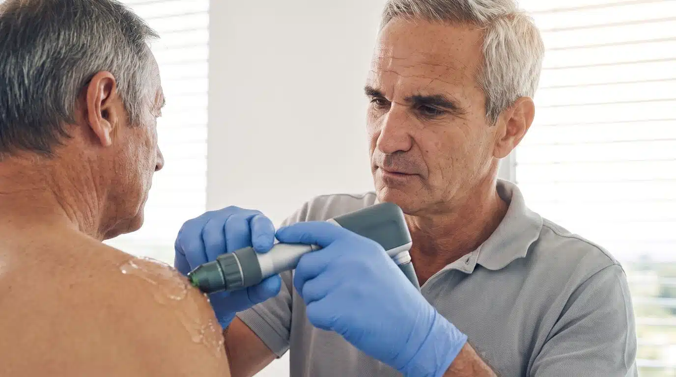
It is important for doctors to explain that calcium deposits diagnosed on an X-ray do not automatically require immediate surgical removal, as many anxious patients often mistakenly fear. Instead, the focus is initially on conservative approaches, such as physical therapy or shock wave therapy, which can be used for certain cases of calcific tendinitis of the shoulder. According to Prof. Dr. Peter Diehl, only in cases of persistent calcium deposits that remain despite conservative measures can targeted, minimally invasive removal or aspiration of the calcium be performed during arthroscopy. Which treatment approach is appropriate in each individual case depends on the size, location, and symptoms associated with the calcium deposit and is always discussed transparently with the patient.
Possible treatment approaches for calcific tendinitis of the shoulder (depending on the indication):
- Comprehensive conservative treatment of the affected area
- Targeted physical therapy for gentle mobilization
- Specific shock wave therapy for the potential dissolution of calcium deposits
- Arthroscopic Removal of Stubborn Calcium Deposits

Shoulder Tendon Tear: When the Rotator Cuff Is Affected
A tendon tear in the shoulder often affects structures that patients commonly associate with the rotator cuff —the group of tendons that surrounds the head of the humerus and provides stability and range of motion. Such a tear can result from acute trauma, but it can also develop gradually as part of degenerative changes. When affected patients receive a diagnosis of such a tear, they often experience understandable uncertainty at first about the course of treatment and the potential limitations they may face in their daily lives.
It is important to make a clear distinction: A tendon tear diagnosed through imaging should not automatically be equated with the need for immediate surgery. The decision regarding treatment must always be made on a case-by-case basis. It is not the mere presence of a structural abnormality that determines the course of treatment—what matters is the patient’s overall situation, including symptoms, function, age, and the demands placed on the shoulder’s strength in daily life. A tear visible on an image is not necessarily the sole cause of current symptoms. Important personal factors such as biological age, individual athletic goals, and occupational demands play a significant role in the medical assessment.
Individualized Treatment Decisions
The appropriate treatment depends on the specific cause and the findings. Not every tendon tear requires surgery—conservative and surgical options must be weighed against one another on a case-by-case basis.

Diagnosis of Shoulder Problems: How to Identify the Cause
According to Prof. Dr. Peter Diehl, the diagnostic process begins with a consultation—or medical history—during which the goal is to document the progression of the symptoms as accurately as possible in order to obtain initial clues about the underlying cause. During this important initial phase, the specialist takes the time to document in detail the exact history of the pain and the patient’s individual limitations. This is followed by a clinical and functional examination: Here, the specialist assesses the shoulder’s actual range of motion, identifies which movements trigger pain, and determines how effectively the muscles can currently stabilize the shoulder.
Possible questions for the medical history:
- Was there a specific accident?
- When did the symptoms begin?
- During which movements do they occur most frequently?
- What types of stress directly trigger the pain?
- What restrictions are currently in place in everyday life?
In addition to the clinical examination, diagnostic imaging plays an important role, particularly magnetic resonance imaging (MRI). It can reveal structural changes such as tendon tears or calcium deposits. However, Prof. Dr. Peter Diehl points out that the patient must lie still during an MRI scan, whereas some symptoms only occur during movement or when the arm is bent at a certain angle. For this reason, MRI findings can never be evaluated in isolation. Only through the judicious combination of all these diagnostic components can a truly complete picture of the underlying condition ultimately be obtained.
Considering Imaging and Function Together
An imaging finding should never be presented in isolation or regarded as the sole truth. Magnetic resonance imaging reveals anatomical or structural changes, while a clinical examination shows how the shoulder actually moves and exactly when symptoms occur. Only by combining both perspectives can a complete picture be obtained: The decision regarding treatment is based on a combination of the patient’s symptoms, the physical examination, and the imaging findings—not on a single piece of the puzzle. This holistic approach effectively prevents premature misdiagnoses and ensures that the subsequent treatment precisely addresses the true cause of the shoulder symptoms.
An MRI image alone does not always fully explain a patient’s symptoms—structure and function must be considered together.
Conservative Treatment Options for Shoulder Pain
The Munich East Orthopedic Center expressly does not view itself as a purely surgical center—conservative treatment approaches are a fundamental part of the center’s mission to spare patients unnecessary surgeries whenever possible and to harness the body’s natural healing powers. Depending on the indication, various methods are used, each of which differs significantly in its mechanism of action and objectives. These include physical therapy, personalized exercise programs, targeted load management in daily life, as well as physical and regenerative therapies such as autologous blood therapy, PRP therapy, and hyaluronic acid treatments.
It is important to note one key limitation: Not every treatment is equally suitable for every shoulder condition. The mere availability of a treatment method should not automatically lead to a recommendation for that specific condition. The decision regarding which procedure is appropriate is always made only after an individual diagnosis, with the treating specialist determining the optimal course of action together with the patient.
Conservative treatments (depending on the indication):
- Physiotherapy
- Personalized Training and Exercise Intensity Management
- Shockwave therapy
- Electromagnetic Transduction Therapy (EMTT)
- Renewable Processes
- Autologous blood or PRP procedures
- Hyaluronic acid
Electromagnetic Transduction Therapy (EMTT)
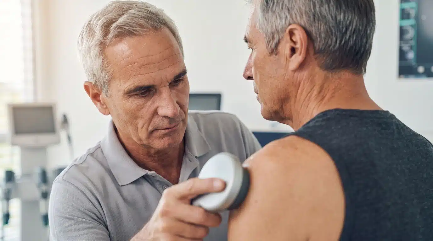
In electromagnetic transduction therapy, a pulsed magnetic field is directed at the area to be treated via a probe. The procedure is intended to influence cells at a deep biological level and promote regenerative or healing-supporting processes. A session lasts about 15 minutes and, in the best-case scenario, is perceived as painless and non-stressful.
However, it is important to set clear limits on expectations: EMTT should not be viewed as a cure-all. It does not heal a torn tendon and does not guarantee a faster recovery. Rather, depending on the diagnosis and treatment goals, electromagnetic transduction therapy can be used as a complementary physical therapy modality within a holistic treatment plan.
Pricing and the number of sessions vary by individual and should be discussed directly with the treatment team.
Shock Wave Therapy for Shoulder Pain
Shock wave therapy is one of the most important conservative or regenerative treatment modalities and is particularly relevant in the context of calcific tendinitis of the shoulder. In this procedure, high-energy pressure waves are directed specifically at the affected area to positively influence problematic calcifications in the tissue. It is also important to note here that no single procedure—such as shock wave therapy versus EMTT—should be presented as fundamentally superior across the board. The appropriate therapy always depends on the patient’s individual diagnosis and personal situation.

When might arthroscopy be recommended?
According to Prof. Dr. Peter Diehl, during arthroscopy, a miniature camera is inserted into the joint through a small incision. The joint is then filled with fluid, allowing the interior of the shoulder to be viewed directly. During the examination, the shoulder can be actively moved to assess, for example, the exact location of a tendon tear. The resulting images are displayed on 4K screens, which allows for a very precise evaluation of the structures.
A key advantage of arthroscopy is that it can be used both for diagnosis and for simultaneous treatment—such as suturing a torn tendon or removing stubborn calcium deposits. This fundamentally distinguishes the procedure from magnetic resonance imaging, which is a noninvasive, purely diagnostic imaging technique. Arthroscopy, on the other hand, is an invasive surgical procedure that allows for direct visualization and, if necessary, immediate treatment of the affected structure.
| Characteristic | Magnetic Resonance Imaging | Arthroscopy |
|---|---|---|
| Type of proceeding | Imaging, non-invasive | Surgical, invasive |
| Patient Position | Lying down, still | Movement is possible during the procedure |
| Purpose | Illustration of Structural Changes | Direct examination and, if necessary, treatment |
| Example | Detection of Tendon Changes | Tendon suturing, removal of calcium deposits |
What is the recovery process like after tendon surgery?
According to Prof. Dr. Peter Diehl, following tendon surgery, the arm can initially be supported or protected in a special pillow for about six weeks. During this phase, the shoulder may initially feel noticeably stiff—mobility must then be gradually and carefully rebuilt. Once mobility has progressed sufficiently under the guidance of a qualified physical therapist, targeted strength training and, later, specific equipment-based training can follow to restore the weakened muscles to full strength. The entire rehabilitation process requires a great deal of patience from the patient and can take up to a year.
One key concept sums up this process: The surgery first restores the anatomy. The body must then relearn strength and coordination. It is important to note that the timeframes mentioned should not be interpreted as fixed recovery periods for every patient—the individual course of recovery can vary significantly depending on the initial findings, age, and the patient’s cooperation during rehabilitation.
Possible treatment pathway:
- Diagnosis
- Treatment Decision
- Surgery, if necessary
- Protection or Recovery Phase
- Mobilization
- Restoring Mobility
- Coordination
- Strength Training
- Equipment Training
- Increase in Workload
- Returning to Everyday Life and Sports
What role does physical therapy play?
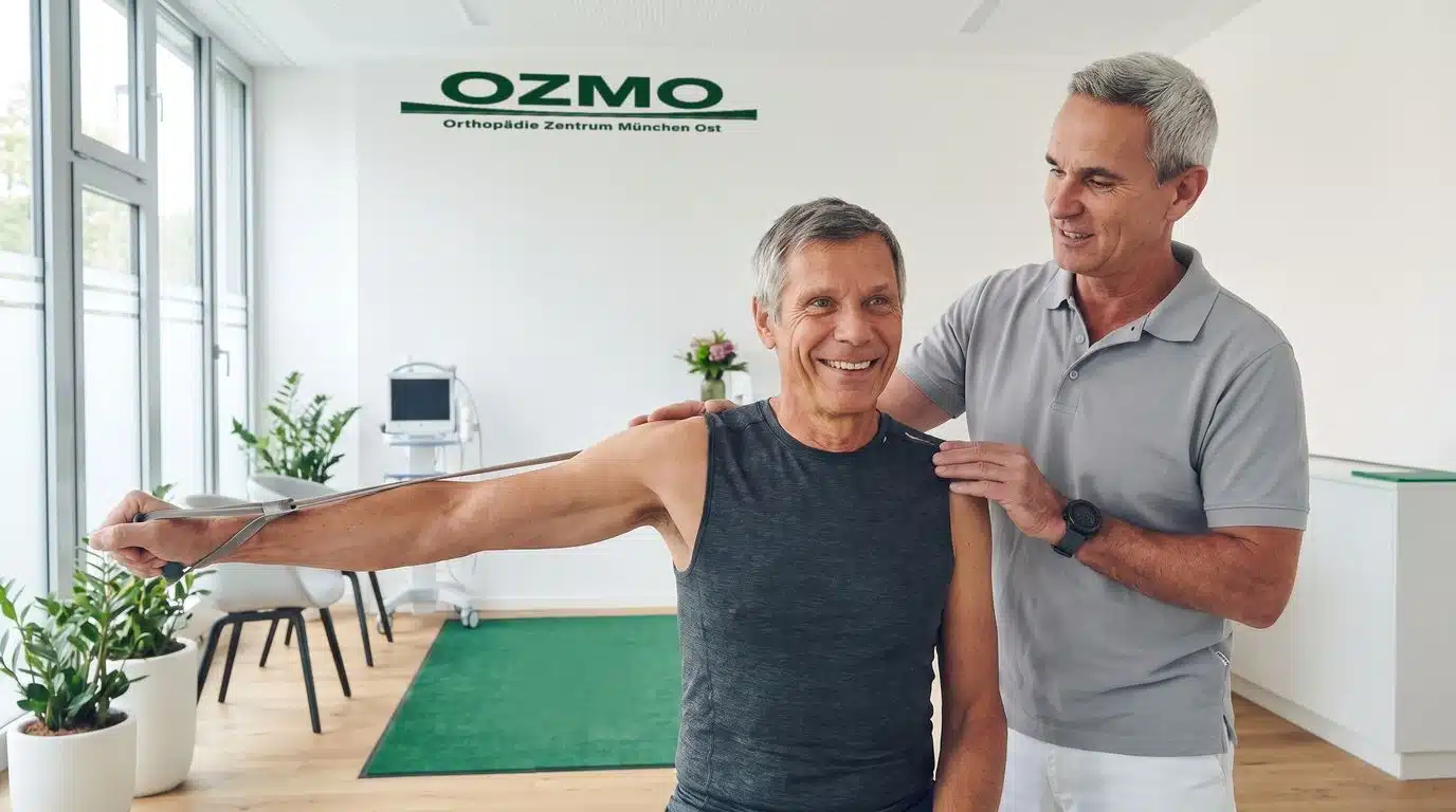
Physical therapy is an absolutely essential and central component of both conservative treatment and post-surgical rehabilitation. Through highly specialized manual techniques and individually tailored exercises, it can play a significant role in bringing muscles and tendons back “into alignment” and stabilizing the shoulder step by step. An important point for all affected patients to note is that well-founded physical therapy plays a role not only after surgery has been completed—it is also very often used as a primary, initial conservative measure to prevent the need for surgery from the outset.
Possible goals of physical therapy:
- Improving Mobility
- Restoration of Functional Movement Patterns
- Improved Muscle Control
- Building Strength
- Improving Coordination
- Support During Rehabilitation
- Returning to Everyday Life and Sports
Whether before a potential surgery, as part of conservative treatment, or following a procedure—physical therapy supports the entire treatment journey and is tailored to each specific phase.
What can you do on your own to help your shoulder?
In an interview, Prof. Dr. Peter Diehl points out that sitting for long periods without taking breaks can be problematic for joint health. This can cause the anterior shoulder girdle to shorten, which can lead to a forward-leaning posture or a hunched back—a posture that many people are familiar with from their daily office routine.
He cites active, regular exercise—particularly rowing—as an important counterbalance: In this movement, the pulling motion is consciously directed backward, thereby targeting precisely those movement patterns that can counteract the forward-leaning posture typical of everyday life. He also notes the benefits of the dynamic back-and-forth arm movement on a cross-trainer. However, it is important to note the following limitation: Individual, primarily preventive exercises should not be viewed as a guaranteed treatment for existing injuries.
Exercise in Everyday Life
Active exercises such as rowing or targeted arm movements on a cross-trainer can help counteract the rounded back caused by prolonged sitting. However, these recommendations are no substitute for an individual medical examination if you have existing symptoms.

When should you see a shoulder specialist?
Persistent or recurring symptoms, pain when raising the arm, nighttime pain, or noticeable limitations in movement should always prompt a prompt evaluation by a specialist. Anyone who ignores these clear warning signs from their own body over an extended period and simply hopes for spontaneous recovery risks an unnecessary worsening of the underlying problem as well as the pain becoming a long-term, chronic condition. Especially if symptoms do not improve over several weeks or even worsen, a thorough diagnosis is advisable to identify the actual cause.
At the Orthopedics Center Munich East, we always follow a personalized diagnostic and treatment plan based on your medical history, a clinical examination, and—if necessary—imaging. During your consultation, the center’s experienced specialists take the time needed to thoroughly analyze your specific symptoms and work with you to find the safest and medically soundest path back to a pain-free shoulder that can once again withstand full activity.
Frequently asked questions
What is shoulder impingement syndrome?
When the arm is raised to the side, structures beneath the acromion may become compressed, which can cause inflammation of the bursa and irritation of the tendon. The exact cause of this compression can vary from person to person.
Does a torn tendon in the shoulder always require surgery?
No, a torn tendon does not automatically require surgery. The decision regarding treatment depends on the patient’s individual situation, including symptoms, age, and shoulder function.
What does an MRI show in cases of shoulder pain?
Magnetic resonance imaging reveals structural changes, such as those in tendons or in the area of the acromion. However, it must always be interpreted in conjunction with the clinical examination and the shoulder’s actual range of motion.
What does a shoulder arthroscopy involve?
A mini-camera is inserted into the fluid-filled joint through a small incision. Treatment can be performed at the same time during the procedure, such as suturing a tendon or removing calcium deposits.
What can I do on my own to help my shoulder?
Active exercise such as rowing and getting enough physical activity in daily life can help support the shoulder. However, if you are experiencing symptoms, it is recommended that you consult a doctor for an individual evaluation.
This information is not a substitute for an individual medical examination. If symptoms persist, it is recommended that you consult a specialist.
Conclusion
Shoulder pain can have various causes—ranging from impingement to calcium deposits to tendon injuries. An accurate diagnosis always combines the patient’s medical history, a clinical examination, and, if necessary, imaging to provide a complete picture of the symptoms.
Depending on the findings, conservative, regenerative, or surgical treatment options may be considered. The goal is always to restore the shoulder’s range of motion, function, and strength on an individual basis—tailored to each patient’s specific situation. Therefore, if you have persistent problems, do not hesitate to seek professional help to prevent the condition from becoming chronic and to find a path to a pain-free shoulder.
Your Path to a Pain-Free Shoulder
Do you have persistent shoulder pain, or would you like to have the cause of your symptoms investigated more thoroughly? Schedule an appointment with the specialists at the Orthopedics Center Munich East.

Tailored diagnostics and treatment – at three locations in the east of Munich. For all insured and private patients.
Memberships & partners



Part of the Ortivity Network · Association doctors of the German Ice Hockey Federation
Clinic Munich
Haar
Münchener Straße 14
85540 Haar
+49 (0) 89 4141 434 – 64
Fax +49 (0) 89 45 45 49 – 39
Opening hours
| Morning | Afternoon | |
|---|---|---|
| Mon | 08:30 – 12:00 | 14:00 – 18:00 |
| Tue | 08:30 – 12:00 | – |
| Wed | 08:30 – 13:00 | – |
| Thu | 08:30 – 12:00 | 14:00 – 18:00 |
| Fri | 08:30 – 12:00 | – |
Appointments by arrangement only
Clinic Munich
Bogenhausen
Denninger Straße 15
81679 Munich
+49 (0) 89 4141 434 – 64
Fax +49 (0) 89 45 45 49 – 39
Opening hours
| Morning | Afternoon | |
|---|---|---|
| Mon | 08:30 – 12:00 | 14:00 – 18:00 |
| Tue | 08:30 – 12:00 | – |
| Wed | – | – |
| Thu | 08:30 – 12:00 | 14:00 – 18:00 |
| Fri | 08:00 – 12:00 | – |
Appointments by arrangement only
Clinic Munich
Feldkirchen
Sonnenstraße 26b
85622 Feldkirchen
+49 (0) 89 90 31 21 4
Fax +49 (0) 89 90 31 52 4
Opening hours
| Morning | Afternoon | |
|---|---|---|
| Mon | 08:00 – 12:00 | 14:00 – 17:00 |
| Tue | 08:00 – 12:00 | – |
| Wed | 08:00 – 12:00 | – |
| Thu | 08:00 – 12:00 | 14:00 – 17:00 |
| Fri | 08:00 – 12:00 | – |
Appointments by arrangement only